What is unique about the Genoray OSCAR Prime Flat Panel Detector C-Arm?
The Genoray OSCAR Prime Flat Panel was launched in 2018, as one of the new models in the OSCAR C-arm

Ultrasound technology is an integral part of today’s medical practice. It is noninvasive, highly accurate, sensitive, and has strong penetration capabilities, thus it is widely used to visualize subsurface conditions for nondestructive evaluation, structural health monitoring, and medical diagnosis.
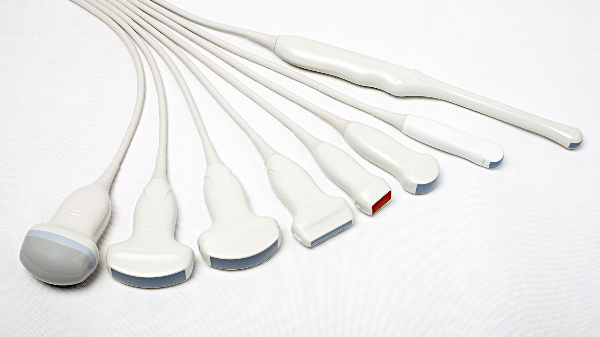
Ultrasound machines use high-frequency sound waves produced by a handheld sensor called a transducer or probe. These transducers contain piezoelectric crystals which vibrate when an electric current is applied. When these crystals vibrate, they transmit ultrasound pulses to organs and tissues being imaged. The probe picks up the reflected sound waves and the crystals turn them back into an electric current. Finally, the machine’s console interprets the signal and converts the electric current into an image seen in a monitor. The shape of the transducers determines its field of view. Transducers are generally described by the size and shape of their footprint.

There are several types of transducers used in medical practice. They operate at different center frequencies, have different physical dimensions, footprints, shapes, and provide different image formats. The scanning depth and resolution are mostly determined by the frequency of the probe. Higher frequencies produce finer images with good resolution. However, because of the shorter wavelength, imaging of deeper structures will be smudgy. Meanwhile, low-frequency probes produce lower resolutions but can reach deeper structures.
The following are commonly used transducers today:

Linear Transducers
Linear transducers generally have a high frequency for better imaging of superficial structures and vessels. The piezoelectric crystals in linear transducers are arranged linearly. This transducer emits a rectangular-shaped beam and produces good near-field resolution images. The frequency, footprint, and applications of linear transducers depend on whether it is designed for 2D or 3D imaging.
Linear transducers for 2D imaging have a central frequency between 2.5 MHz and 12 MHz. These transducers have a wide footprint and keep the same field of view at the deep part. They are typically used for breast examinations, body fat and muscle measurement, thyroid evaluation, and blood vessel visualization. Linear transducers used for 3D imaging have a central frequency between 7.5 MHz and 11 MHz. They also have a wide footprint and are commonly used for breast, thyroid, and vascular applications.

Convex Transducers
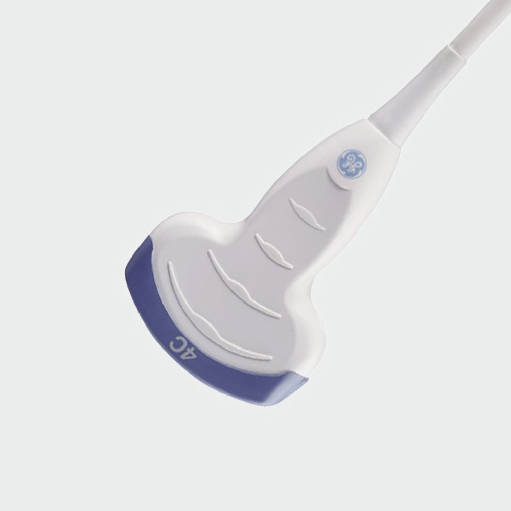
These probes have a widened footprint and lower frequency for transabdominal imaging and a widened field of view. They have piezoelectric crystals arranged in a curvilinear fashion which produces a convex-shaped ultrasound beam. This type of transducer is ideal for in-depth imaging, even when resolutions decrease with depth. Footprint, frequency, and applications of these transducers depend on whether they are designed for 2D or 3D imaging.
Convex transducers used for 2D imaging has a wide footprint with a field view spread at the deep part. It has a central frequency between 2.5 MHz and 7.5 MHz. They are typically used for abdominal and organ examinations. Convex transducers used for 3D imaging have a wide footprint probe head ideal for scanning deeper structures. They have a central frequency between 3.5 MHz and 6.5 MHz. They are generally used for abdominal examinations.

Phased Array Transducers
These transducers have a piezoelectric crystal arrangement known as phased-array. They have a small footprint resulting in a narrow beam point with a widely spread field of view at the deep part. They operate at a low frequency between 2 MHz and 7.5 MHz with a low near field resolution. Several applications include abdominal, cardiac, and brain examinations.

Endocavitary Transducers
These transducers allow internal examinations of the patient. They are designed to enter and fit in specific body orifices like the vagina and rectum. They have a small footprint and operate at a frequency range between 3.5 MHz and 11.5 MHz.

Transesophageal Transducers
These transducers are designed for internal imaging and function similar to flexible endoscopes, with controls on the probe body that allow the technician to manipulate the transducer lens inside the patient. These transducers have a small footprint and are used for internal examinations. They can be used to get a better image of the heart and its surrounding tissues through the esophagus. These transducers operate between 3 MHz and 10 MHz.
Always return the handpiece to the nonconductive holster or quiver when not in use. Place the electrode tip in its insulated container while on the surgical field.
Identify the correct plug ports for connecting the appropriate cables. Do not use sharp or metallic instruments to attach the electrosurgical unit cables to drapes. This can damage electrical cables, cause electric shock to the patient or operator, or ignite drapes.
Most importantly, keep the equipment well-maintained and tested to ensure they are working properly.

Related Posts
The Genoray OSCAR Prime Flat Panel was launched in 2018, as one of the new models in the OSCAR C-arm

What is an ultrasound transducer? Ultrasound technology is an integral part of today’s medical practice. It is noninvasive, highly accurate,

What is LigaSure technology and how does it work? LigaSure technology is considered as the standard in vessel sealing. In
Headquarters
685 Orchard Street, New Bedford MA 02744
Phone
508-996-9005
Email
info@MED.equipment